Focus shifts to device fragments, small miscellaneous items in RSIs


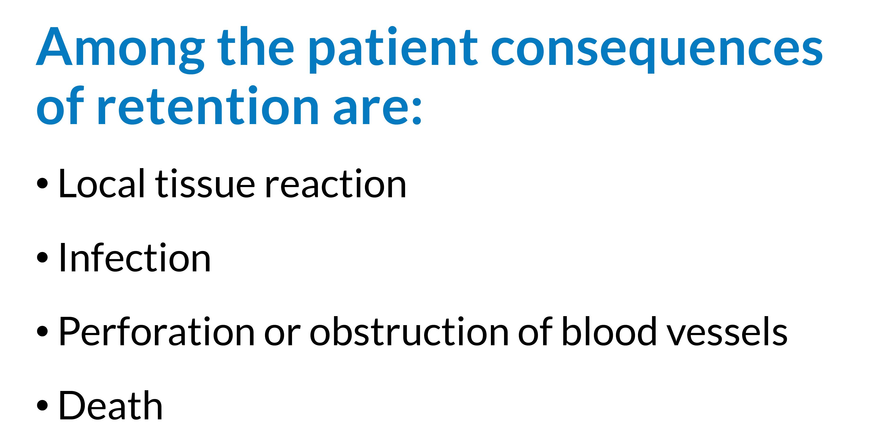
Though retained surgical items (RSIs) cases are rare, they do happen, and they take a heavy toll throughout the system in terms of steep fines, malpractice claims, and compromised patient safety. Estimates of RSIs range from 1 in 1,000 to 1 in 7,000 procedures. And a 2003 study by the Agency for Healthcare Research and Quality found that patients with RSIs had a mortality rate 2.14% higher than controls, excess hospital stays of 2.08 days, and excess costs of $13,315.
There is no national reporting system for RSIs, but state and federal agencies along with accreditation organizations have recommended action to prevent such events.
RSIs are considered a serious reportable event (SRE) by the National Quality Forum and a sentinel event by The Joint Commission. The Centers for Medicare and Medicaid Services lists RSIs among the hospital-acquired conditions for which it will no longer provide payment under the Inpatient Prospective Payment System.
The state of California in 2007 began mandating that hospitals report cases of RSIs and other SREs and levying administrative penalties in cases where serious harm has occurred (www.cdph.ca.gov/Pages/NR13-005.aspx).
In February 2013, the California Department of Public Health (CDPH) issued penalties against 2 hospitals for RSI cases:
- One hospital was fined $100,000 for failing to develop and implement a surgical count policy and procedure specifying that small items would be accounted for prior to closure. A Raney clip was left inside a patient’s skull after brain surgery.
- Another hospital was fined $75,000 for leaving behind a stiffener stylet (guide wire) from a Groshong catheter.
Interestingly, these fines were for retained small miscellaneous items and device fragments.


The variety of small miscellaneous items and unretrieved device fragments (UDFs) that are being left in patients has increased, and they are gaining attention, says Verna Gibbs, MD, who developed the NoThing Left Behind® project for RSI prevention (www.nothing leftbehind.org).
Dr Gibbs has termed these items “surgical junk.”
Orthopedic surgery cases account for the largest number of these small miscellaneous items and UDFs, which often are the result of breakage of tools when used against bone. New to the scene are items such as guide wires, catheters, stents, and sheaths, which are left in patients whose procedures are performed in cardiac catheterization labs, interventional radiology labs, and hybrid ORs.
For example, problems can occur when a guide wire gets tangled around a stent and the wire fractures, leaving behind a small part of the wire, or a subcutaneously placed catheter snaps upon removal and part of it is left interstitially.
Device fragments can result from instrument failure that develops from extensive use (burrs, loose parts) or faults in new instruments, such as poor welds or rough surfaces.


In 2008, the Food and Drug (FDA) Administration issued a medical device safety alert warning of serious adverse events associated with UDFs and provided recommendations to mitigate these events (www.cdph.ca.gov/Pages/NR13-005.aspx).
The FDA characterized a UDF as “a fragment of a medical device that has separated unintentionally and remains in the patient after the procedure.”
The FDA’s Center for Devices and Radiologic Health (CDRH) receives nearly 1,000 adverse event reports each year related to these items.

RSI-related safety notices on the FDA website reflect a range of consequences. For example, in 1 safety notice, the FDA describes the fracture of the distal tip of an epicardial pacing lead. The tip was left in the patient without any adverse consequences (www.fda.gov/MedicalDevices/Safety/AlertsandNotices/TipsandArticlesonDeviceSafety/ucm203731.htm). In another instance, a fractured guide wire lodged in a coronary artery during a cardiac catheterization, resulting in the patient’s death from cardiac tamponade (www.fda.gov/MedicalDevices/Safety/AlertsandNotices/TipsandArticlesonDeviceSafety/ucm070187.htm).
In addition, during MRI procedures, magnetic fields may cause metallic fragments to migrate, and radiofrequency fields may cause them to heat, which can lead to internal tissue damage and burns.
“What’s important to point out about these miscellaneous small items and unretrieved device fragments is that physicians may not think they need to disclose to patients that they have left something behind if they decide not to take them out,” says Dr Gibbs, professor of clinical surgery at UCSF and a surgeon at the San Francisco Veterans Affairs Medical Center.
“This is bad practice,” she says. “Physicians have a moral and ethical obligation to tell the patient. It is important for the patient to know in case an MRI is needed in the future.”


A 2012 study led by Susan Moffatt-Bruce, MD, PhD, chief quality and patient safety officer and associate professor of surgery at Ohio State University Wexner Medical Center in Columbus, examined risk factors for intravascular retained small miscellaneous items and device fragments.
The retrospective study of 83 RSIs found that 13 cases involved intravascular small miscellaneous items and device fragments—8 guide wires, 4 catheter/catheter fragments, and 1 coil.
Locations included:
- 3 catheter fragments were retained in the pulmonary arterial tree. Of those, 1 broken catheter tip embolized into a distal pulmonary arterial branch, and 2 catheter fragments were located in the heart.
- 1 guide wire was retained in the subclavian vein
- 2 guide wires were extending between the heart and iliac vein
- 2 guide wires were extending between the inferior vena cava and the right atrium
- 3 guide wires were extending between the superior vena cava and the inferior vena cava
- 1 catheter and the coil were located peripherally in smaller vessels.
Procedural factors significantly associated with the retained intravascular items included:
- Technically difficult procedure—a procedure that did not proceed as planned (ie, took more than 1 attempt)
- Unfamiliarity with the equipment—new equipment or equipment that did not work as expected or malfunctioned
- Difficult/emergent setting—a procedure done emergently, often without enough time to go through the usual safety steps, in a less than optimal environment.
“One of the things we found was that radiology under-reads, or the item is missed on the initial read in a significant number of cases,” says Dr Moffatt-Bruce. Seven of the 13 items were missed on confirmatory postprocedural x-rays. Most of the retained items were found within 48 hours and were removed the same day. They were detected by means of interventional radiology procedures. Some items, however, were indwelling up to 6 weeks before being identified.
“Though all of the patients did well, it is a significant patient dissatisfier to have to undergo another invasive procedure,” she says.
Strict adherence to protocols and stringent radiographic review, along with standardized team training, checklists, and documentation, are needed to prevent these incidents.

Another of the penalties issued by CDPH in February 2013 was a fine of $100,000 for a surgical lap pad left in a patient despite the use of a 2-D matrix computer sponge counting device.
Stanislaw Stawicki, MD, and Dr Moffatt-Bruce led a multicenter study to gain a better understanding of why RSIs continue to occur, despite the use of radiofrequency tagged sponges and wand systems and mandatory x-rays, and how to reduce such events.
One of the analyses combined data from 2 previously published case-controlled studies on RSIs (Gawande et al, 2003; Lincourt et al, 2007) with data from their study on 59 RSIs and 118 matched controls from 5 institutions gathered over a 6-year period.
“Many variables that were not significantly associated with RSIs when data from the 2 previous studies were combined became significant when our data was added,” says Dr Stawicki, director of research for the division of trauma/critical care and associate professor of surgery at Ohio State University Wexner Medical Center.
Results showed that counts had been documented as correct in 45 of 59 cases, even though a sponge was later found inside the patient. In 13 of 27 cases an RSI was missed on initial confirmatory x-rays. In 2 of 32 cases in which radiofrequency tagging systems were used, RSIs were missed.
The biggest risk, says Dr Moffatt-Bruce, is that humans are doing the operating and humans are doing the counting. “Our goal is to minimize that risk as much as possible through the adherence to and enforcement of a standardized process for counting,” she says.
“All technology does is layer on another practice,” notes Dr Gibbs. “Humans are making errors in counting without technology, and humans will continue to make mistakes with technology added.”
Dr Gibbs has been testing and teaching a simplified, transparent standardized manual sponge management practice, called Sponge ACCOUNTing System, which she says is being used in hundreds of hospitals across the country. The practice requires the nurses to manage the sponges only in multiples of 10 and to account for them at the end of the case rather than just count them.

Each member of the OR team plays a part in minimizing the risk of losing small miscellaneous items and generating UDFs, says Dr Gibbs.
- Surgical technologists should inspect any device before handing it to the surgeon and again when it is passed back after use.
- Surgeons should perform a methodical wound exam before closing every wound.
- Circulating nurses should direct the activities to account for all 4 classes of surgical items—soft goods (eg, sponges, towels), sharps (eg, needles, blades), instruments, and small miscellaneous items and device fragments.
Dr Gibbs says she would like to see a checkbox added to nursing operative records to signify a correct count of all small miscellaneous items. Currently nurses are bundling small miscellaneous items either with sharps or instrument counts.
In cath labs and procedural areas, proceduralists have to develop and adopt practices to account for all surgical items at the end of the procedure. One example would be to have a memory aid added to the central line-associated bloodstream infection (CLABSI) procedure list to visually confirm that the guide wire is in the kit at the end of the insertion. This would ensure that it wasn’t inadvertently left in the patient.
“Instead of trying to assess the risk of an RSI based on the type of case or characteristics of the patient,” says Dr Gibbs, “it would be better to look at the risk of the providers performing and assisting with the surgical procedures. The risk is in the personnel and the environment in which they work.
“An RSI means the OR team has poor practices and is not working together. I call an RSI case a canary in the surgical coal mine,” she says.
A lot has been learned from wrong-site surgery, says Dr Gibbs. By implementing timeouts and using checklists, OR staff began to standardize practice, communicate, and work together. Similar systems are needed for prevention of RSIs.
Dr Moffatt-Bruce notes that because of their study, Ohio State University Wexner Medical Center invested in team training through crew resource management. Some 38,000 staff have been trained.
Crew resource management speaks to the basic premise of sharing the same mental model in the OR, whether it’s during the procedure or during the counts, she says. The model makes the surgeon responsible for doing the timeout as well as the debriefing at the end of the case to ensure nothing is left behind.
References
Ergova N N, Moskowitz A, Gelijns A, et al. Managing the prevention of retained surgical instruments: What is the value of counting? Ann Surg. 2008;247:13-18.
Gawande A A, Studdert D M, Orav E J. Risk factors for retained instruments and sponges after surgery. N Engl J Med. 2003;348:229-235.
Gibbs V. A surgical junkyard. CHPSO Patient Safety News. 2011;3:1.
Lincourt A, Harrell A, Cristiano J, et al. Retained foreign bodies after surgery. J Surg Res. 2007;138:170-174.
Mehtsum W T, Ibrahim A M, Diener-West M, et al. Surgical never events in the United States. Surgery. 2013; 153:465-472.
Moffatt-Bruce S D, Ellison E C, Anderson H L, et al. Intravascular retained surgical items: A multicenter study of risk factors. J Surg Res. 2012;178:519-523.
Rowlands A, Steeves R. Incorrect surgical counts: A qualitative analysis. AORN J. 2010;92:410-419.
Stawicki S P A, Moffatt-Bruce S D, Ahmed H M, et al. Retained surgical items: A problem yet to be solved. J Am Coll Surg. 2013;216:15-22.
Zhan C, Miller M R. Excess length of stay, charges, and mortality attributable to medical injuries during hospitalization. JAMA. 2003;290:1868-1874.